Cardiac MRI
Why CARDIAC MRI?
1. Superior field of view than 2D-echo
2. Functional information derived including global LV and RV volumes and mass, can be applied to all sizes and shapes and shape of ventricles, even those with remodeling
3. Volume and mass assessment with cine imaging, identification and quantification of fibrosis, quantification of valvular lesions- highly accurate and reproducible
4. Right Ventricle assessment (3-dimensional nature)
5. Accurate identification of even subtle regional wall motion abnormalities with the use of steady-state free-precession sequences, which provide excellent delineation of the blood–myocardium interface
6. Flow assessment- velocity encoded CMR sequences, which enable accurate quantitative measurements for stenosis (peak velocity and by applying the Bernoulli equation peak gradient) and regurgitant valvular lesions (regurgitant volume and fraction)
7. In suspected shunt, the pulmonary-to-systemic flow ratio (Qp/Qs) can be determined by measuring flow in the main pulmonary artery and the ascending aorta
8. Myocardial tissue characterization
9. Intracardiac thrombi detected by early imaging (1-3min) &
images acquired late after 5-20min- Late gadolinium enhancement Cardiac MRI
Indications
1 Ischemic Cardiomyopathy - Viability Imaging
2 Myocarditis
3 Non-ischemic Cardiomyopathies: Hypertrophic Cardiomyopathy (HCM/HOCM), Dilated Cardiomyopathy (DCM), Arrhythmogenic right ventricular cardiomyopathy (ARVC), Restrictive CM, Secondary to tuberculosis
4 Infiltrative – Amyloidosis, Sarcoidosis, Anderson-Fabry ds,
5 Iron Overload Cardiomyopathy –THALASSEMIA T2* imaging
6 Constrictive Pericarditis
7 Non-compaction Syndrome
8 Cardiac Masses
9 Valvular heart disease
10 Congenital heart disease
11 Other Cardiomyopathies (Tako-Tsubo, Churg-Strauss etc.)
 |
|

 MYOCARDITIS MYOCARDITIS
|
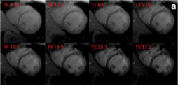 T2* Imaging Iron overload T2* Imaging Iron overload
|
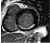
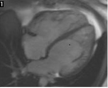
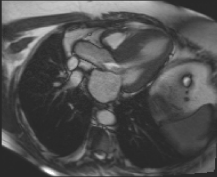 Hypertrophic Cardiomyopathy with SAM and accelerated flow Hypertrophic Cardiomyopathy with SAM and accelerated flow
|
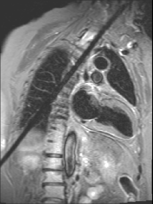
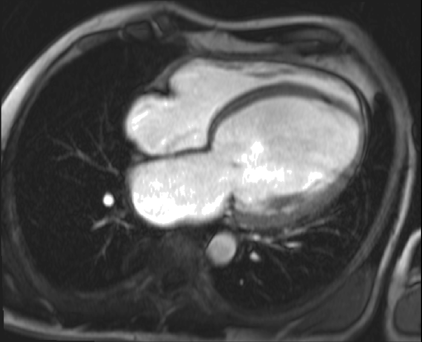 Dilated Cardiomyopathy Dilated Cardiomyopathy
|
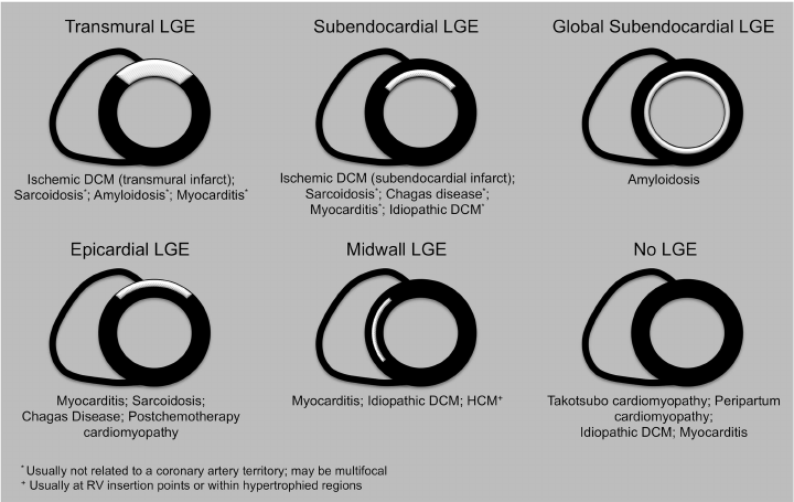 PATTERNS OF CONTRAST ENHANCEMENT PATTERNS OF CONTRAST ENHANCEMENT
|
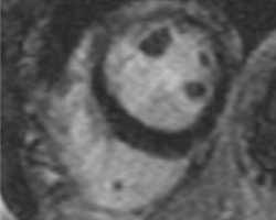
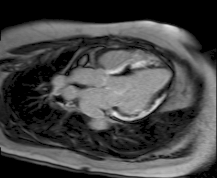 Sarcoidosis Sarcoidosis
|
 Non Compaction Syndrome Non Compaction Syndrome
|
 Pericarditis Pericarditis
|
Contraindications & Limitations
1 Implanted metal device (e.g. pacemakers or defibrillators, cochlear implants, cerebral aneurysm clips), or who may have iron fragments in their eyes
2 Orthopaedic pins, mediastinal clips, coronary stents, and the majority of artificial heart valves are safe to scan
3 In patients with very fast heart rates (tachycardia), or frequent irregular beats (arrhythmias/ectopic beats), ECG gating can prove unreliable
4 Patients with severe heart failure may struggle to lie flat for the duration of the test
5 There are no known risks from undergoing MRI during pregnancy. However, as with any medical investigation, a patient who suspects that she is pregnant should seek advice on the risks versus benefits of undergoing the investigation
6 Few will suffer from claustrophobia to a degree that will not allow them to tolerate a MRI scan
7 Very obese patients may not be able to fit comfortably within the MRI machine
 Interventional Radiology
©2018 Nirman Diagnostics.. All rights reserved.